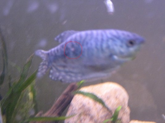
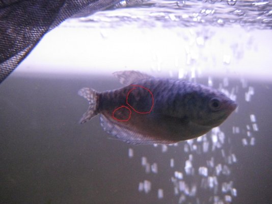
Mine is still acting normal, fins are healed, but the reddish spot that became a 'C' shape, is now a small, raised circle. Becoming more raised, can't tell if it's fuzzy, or scabbed. . There's also 2 other new spots where it looks like a single scale is barely attached, but no discolouration.
I was looking into that bacteria, and it was actually kind of difficult to google decent info relevant to fishkeeping. I found this, which is similar, but mine doesn't have all the symptoms.
In the early stages, scales protrude in patches (otherwise fish look healthy). As the disease progresses, scales begin to stick up all over the body. This is caused by the formation of small bubbles filled with serous fluid (pustules). These pustules are situated under the skin in places where scales are attached to it. Sometimes loss of scales will occur. The formation of pustules, scale protrusion, and loss of scales cause the disruption of gas exchange, which is particularly dangerous for young fish whose gill apparatus is not well developed and for whom dermal respiration is very important. Diagnosis cannot be based solely on these symptoms, as scale protrusion is observed in fish infected with Mycobacteriosis and Ichthyosporidium. It is necessary to carry out microscopic examination of smears of the contents of the pustules and tissues adjacent to them. It might be necessary to perform microbiological examinations of inoculations of the hypoderm and viscera (the kidneys, liver, heart, and spleen).
otherwise known as infectious scale protrusion, is a common aquarium fish disease. It is thought to be caused by the bacteria Aeromonas punctata and Pseudomonas fluorescens. The condition slowly reaches epizootic proportions and is often fatal. The bacteria which cause the disease are widely spread in nature and are brought into aquaria from natural reservoirs. Disease can be transmitted by infected fish which have not been quarantined before introduction into the aquarium as well as through water, gravel, and plants from infected tanks. Fish can also become infected through the use of nets and other equipment used in several tanks.
How to cure:
The disease can only be treated if caught in the early stages, when scales stick up in patches. Antibiotics Bicillin -5, Biomycin or Sulfanilamide (white Streptocide) are used to treat scale shedding. Basic Violet K can be used if fish are treated in a separate tank. Fish that show extensive scale protrusion should be disposed of. Disinfect the aquarium, fishing tackle and other equipment with 5% solutions of HCl (Hydrochloric acid) and H2SO4 (Sulphuric acid) or Chloramine. Gravel should be boiled or tempered as a means of disinfecion. Plants are treated with Bicillin-5 solution
It also has a few symptoms similar to TB, but there's far more symptoms that aren't apparent. Never dealt with either of these.
I'm going to lfs tomorrow to get a lid for the QT, then I'm putting him in. Any thoughts on what medication I should at least try? Since Melafix antibacterial seems to have no effect. None of the ones listed are available here.