Please ID Hitchhikers

The friendliest place on the web for anyone with an interest in aquariums or fish keeping!
If you have answers, please help by responding to the unanswered posts.

Ducaroo

Aquarium Advice Apprentice
Joined
Nov 16, 2008
Messages
10
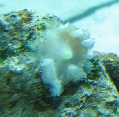
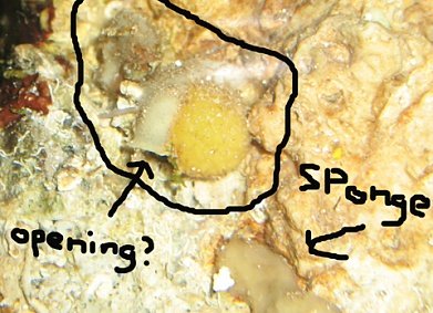
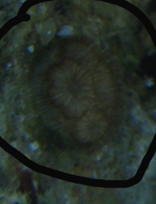
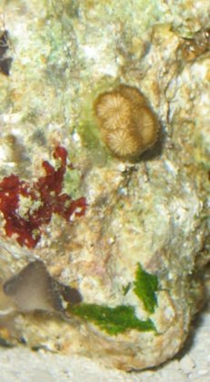
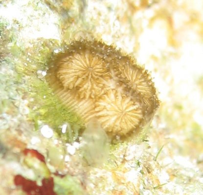
I have a 70gal tank and cycling LR for 3 weeks on one rock. First photo is some kind of anemone? about the size of a half dollar, pink base, likes low light, flattens w/ bright lights and tentacles become inflated. The second photo is some growth about 1/4 inch wide on side of same LR. Third photo is from bottom of LR. It looks like a yellow ball with spikes on it, and attached to the side is a white tubular thing with maybe an opening on the large end of the opening(not sure). Any help would be appreciated. Do any of these have to be ejected?
 

Attachments

  • Ann.jpg
    Ann.jpg
    59 KB · Views: 72
  • HBV.jpg
    HBV.jpg
    71.1 KB · Views: 62
  • Coral.jpg
    Coral.jpg
    33.6 KB · Views: 89
Not sure, but I'll take a stab to get the ball rolling!

Upper left looks kind of like a really unhappy, bleached bubble tip anemone? Or at least an unhappy anemone of some type. Not sure on that one...

Upper right is a collection of sponges and tunicates. All good things to have.

Lower shot... really hard to tell if that's living tissue or just a dead coral skeleton. Can you see any tissue, or does it inflate/expand at all during the day? How big across is the largest circle?
 
I believe the 1st pic is a rock anemone, not a happy one either. I agree with the 2nd pic and the 3rd I can't tell what it is. Can you get a pic of it with your flash so we can see it better?
 
Holy Tuna Cakes! I had no idea there was such an animal as a tunicate. You guys are great! Attached is hopefully a better picture of the coral(?). It's about 3/8 inch in diameter. I cannot tell if it's been huffing and puffing during the day, but it seems to be getting bigger, slowly. Thanks again for your diagnosis :D
 

Attachments

  • IMG_3700.jpg
    IMG_3700.jpg
    49.8 KB · Views: 86
Hmm... just can't tell from that small of a picture. Kinda looks like it could be a type of button polyp... a palythoa of some type? Maybe????

Does it have a tiny little slit in the middle that looks like a mouth?
 
Kurt you may be on to something there.

Are each of the "circles" individual polyps? The pic is so small it's really hard to tell what it is. When you upload your image try and keep it at 800x600.
 
I just can't tell if the "stripes" I'm seeing is the coloration of the tissue (like a lot of palythoas) or is actually skeleton. That first picture looks like skeleton, but this last picture looks more like living tissue.

Ducaroo... if you GENTLY poke it with a stick or something (NOT your hands... palythoas are very toxic) does it close up at all?
 
Ok, here's another shot of object #3. I tapped it gently. It was hard to the touch. It responded by changing color from brownish-yellow to white and posting a "This coral not in foreclosure. Trespassers will be stung" sign. Each circle from the picture does appear to be an individual whatever. The stripes look like it's part of the skeleton(?). Thanks for your input!
 

Attachments

  • IMG_3722.jpg
    IMG_3722.jpg
    68.6 KB · Views: 67
Okay... scratch palythoa. Sounds like a hard coral of some sort, but I haven't got the foggiest. Kind of looks like a little colony of baby fungias, but that thing they're growing out of is kind of throwing me for a loop. Interesting hitchiker!
 
That is one of the wildest looking things I've ever seen. I have no idea what the heck it is. If you don't mind I'm going to forward a copy of that pic to a couple reef buddies I have that are pretty good at figuring things out or at least pointing me in the right direction. My curiosity is going nuts now.
 
I found a picture in Veron's coral book that looks dang near exact to that last picture (complete with the striped looking base)... except it describes it as a solitary coral that lives on substrate - kind of like a fungia. Perhaps they start as colonies like fungia?

Anyway... the coral I'm looking at is a Heterocyathus aequicostatus, part of the Carylphylliidae family. I did a quick look on the web for a picture, but couldn't really find one. Don't really feel like scanning a copyrighted picture and posting it, so you may do a little Googling with that name and see if you can find anything.
 
Wow! You guys are amazing:D. I did a search for Leptastrea sp., then did a search for Heterocyathus aequicostatus. I found the Heterocyathus aequicostatus at a website, but while scrolling through the other photos, I found a pic of a specimen that looks very much like mine, but bigger. It's labeled as Pseudosiderastrea tayami of the Siderastreidae family. Of course, being a newbie, it's all Latin to me. What is it? What would be a good reference for a newbie to get a handle on the classification, morphology, etc. of corals?:confused:
 
You're probably the better judge of what you have, well... since you have it... but Pseudosiderastrea seems more colonial to me. It's a colony of polyps that share the walls of their skeleton. Your pictures seem to be a group of individual polyps - each one its own thing.

The coral I noted is part of the Caryophyllidae Family. Another related to that is Caryophyllia... you might Google Image that name and I think you'll find a bunch similiar to your pictures. There's also Phyllangia... but that appears to be an Atlantic species and not a Pacific. I'm assuming your rock came from the South Pacific?
 
It was also suggested to me that it could be a type of Blastomussa that is very unhappy. I have blastos and I'm not so sure I agree with that assessment.

Kurt this is what I found for the coral you listed. Doesn't really look like it to me. This coral looks more like the way a bubble coral might grow. Anyway here is the link to it.

Heterocyathus Aequicostatus

While searching and looking at pics I found this to be a possibility as well. Keep in mind that your coral is stressed and bleached and working on coming back so don't focus on the color. It's possible it is Galaxea fascicularis The polyp structure is similar to some extent. I would photograph the coral you have every couple of days to document the changes it goes through.

I agree with Kurt that it's probably not Pseudosiderastrea tayami, each poly shares a wall as he mentioned and yours have clearly defined polyp structure. I think as the coral gets happy again an shows some color we might be able to figure out what it is. It was also suggeted to me that you present it with some mysis to see if it reacts by extending feeder tentacles. Just a thought...I'm still looking.....
 
Back
Top Bottom